Patient comfort and safety

In 3D X-ray imaging patient should stay still for the whole duration of the scan in order the machine to acquire high quality data. Nowadays the 3D X-ray units take hundreds of projections and the scan takes a while. Also, the radiation from X-rays is harmful to the patient, so there is a health risk, too. Still, 3D X-ray is becoming more and more popular, because it's a great way for doctors to give accurate diagnosis.
The whole medical field is seeking ways to lower the patient dose and make the scans faster. Te solution could be the reconstruction process and how to change it. Reconstruction process transforms the imaged 2D X-ray projections into a volume information, which is often viewed as cross-section slice images and shifting between them.
To significantly lower the patient dose and making the scans faster is the ultimate way to enhance the 3D X-ray imaging for patients and hospitals alike. Taking fewer projections is a straightforward way to cut down the patient dose. This also speeds up the scanning procedure accordingly. The challenge is how to cope with less data. Eigenor reconstruction software can survive with dramatically fewer projections and still deliver high quality 3D data. Decreasing the patient dose by 50% is possible and simultaneously also speed up the scanning procedure. Serve your customers with healthier imaging and plesent process.
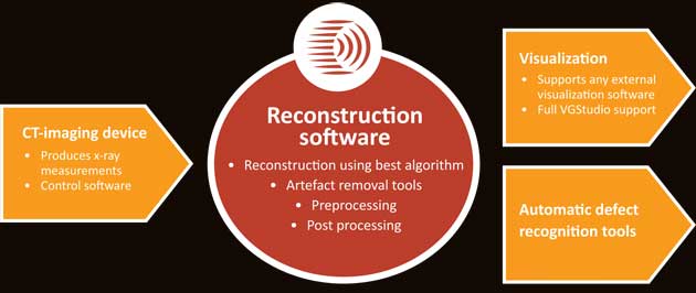
Software solution for you
Whether you're a X-ray machine manufacturer or an end user of one device, our solution is available for you. Eigenor software can be integrated into any existing 3D X-ray system. The software is hardware independent and requires just the projection data to work. The visualisation of the volume data stays the same as all the other operations and you can continue to your work as usual with the enhanced system. High quality data with faster scans and less dose for the patient meaning also more patients per day that the system could handle before. Exploit the full potential of your 3D X-ray unit.


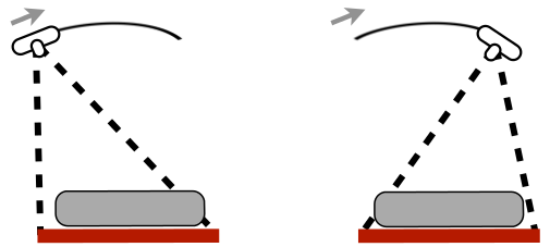
Left: Cross section slice image with 400 projections
Right: Eigenor reconstruction cross section slices fron only 200 projections, i.e. 50% less patient dose.
Machine manufacturers can focus on the hardware development and design. Companies like Planmeca have chosen to deploy Eigenor reconstruction software into their 3D medical units. Eigenor software is integrated into the system and takes care of the reconstruction process of the data and forwards the results onwards, eventually to be visuallised to the user, doctors and X-ray unit operators. You can have significant savings in R&D costs with ready software solution and rapidly develop a world class 3D X-ray machine. Many machine manufacturers have found our simulation services really useful during early design phase to verify reconstruction results before any machine prototype exists.
Transforming 2D medical units into 3D
There are lots of medical units where the X-ray source can move in limited angle around the patient. These kind of 2D units are relatively cheap compared to full 3D CT units and can provide a bit better view than static 2D units. The ability to move the source in limited angle combined with Eigenor software opens up totally new possibilities and markets. Proving results that are usually only received with full CT unit can be now achieved with limited angle measurements.
Three reconstruction methods to fullfil your application needs
Variety of medical scanners and their requirements has driven our software development. Broad selection of different reconstruction methods satisfies special requirements of different applications and allows us to serve you with the best fit for your unit. Eigenor reconsutrction software supports all typical scanning geometries and beam types.
Eigenor reconstruction methods available as software
Eigenor SART: SART is an iterative method more robust with noise and capable of providing results from limited angle measurements also. Support also larger set of different measurement geometries than FDK and thus very good algorithm for experimental CT measurements. Eigenor SART implementation is fully GPU accelerated.
Eigenor PSIG: PSIG is intended for more complex measurement cases, where none of the traditional algorithms typically yield any meaningful results. With PSIG you can expand the possibilities of CT beyond what you previously considered as limits of CT.
Eigenor FDK: Traditional backprojection method completes the reconstruction method portfolio of Eigenor. FDK is the best method for the tradiational CT units, excelling in measurement scenarios where hundreds of projections are available and X-ray source-detector pair movement trajectory is circular. Also FDK requires measurements from full angle, minimum of 180 degrees, most commonly a full circle. Eigenor FDK is optimized for GPU usage to provide reconstruction results fast.
See hardware recommendations for each of the methods.